My Years in Neurofibromatosis Research: Inner Reflections
Karin Soares Cunha, PhD, Professor of the Department of Pathology, School of Medicine, Fluminense Federal University, Brazil
Discover more articles in the Women in NF series by clicking here.
I am honored to join this tribute to women’s essential work in the NF community, emphasizing the importance of diverse perspectives in advancing the understanding and management of these complex diseases. This initiative is crucial, especially in a world where many women still face challenges in claiming their rightful place.
From Frustration to Motivation: My Neurofibromatosis Journey

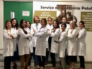
Dr. Cunha’s team 2018
My NF research journey began in 1999 during my specialization in stomatology at the Federal University of Rio de Janeiro, Brazil. That year, a six-year-old boy with a facial plexiform neurofibroma (pNF) visited our clinic. Like many NF1-affected families, his mother was seeking both diagnosis and effective treatment for her son. Faced with an inoperable tumor and no drug treatments available, this first encounter with NF1 motivated my entry into NF research.
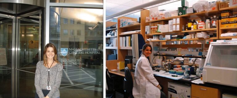
During my Master’s in Pathology in 2000, I began researching neurofibromas’ pathogenesis, continuing this focus throughout my Ph.D. in Pathology. In 2010, I enhanced my expertise in the NF field through training at Harvard University with Dr. Scott Plotkin’s team, including Dr. Anat Stemmer-Rachamimov and Dr. Fabio Nunes.
Upon returning to Brazil, in a professor position, I established the Neurofibromatosis Study Center at Fluminense Federal University. In partnership with Dr. Mauro Geller, we have joined forces with the National Center for Neurofibromatosis, expanding our capacity to manage and research NF1 despite the significant barriers posed by Brazil’s limited science investment. Within the center, I am pleased to have encouraged and participated as a mentor in the training of over 30 students in scientific initiation, master’s, and doctoral programs in the NF1 field, with the majority being young women.
Some Personal Contributions to NF1 Research
Neurofibromas and Hormones
One of the focuses of my research has been the effects of hormones on the pathogenesis of neurofibromas. At the beginning of my career, scientific research into this topic was essentially unexplored despite long-standing clinical evidence indicating the impact of puberty and pregnancy on the initiation and growth of cutaneous (cNFs) and subcutaneous neurofibromas (scNFs), as well as the influence of pregnancy on pNFs, with reports of rapid growth and malignant transformation.
Over the years, our research group,1,2, along with others, three, has demonstrated that the progesterone receptor (PR) is frequently expressed in cNFs and pNFs, while only a few tumors express the estrogen receptor (ER). Our group has also revealed that cNFs commonly express the non-classical receptor for estrogen, GPER. Additionally, we showed that PR and GPER expression positively correlates with the cell proliferation index in human tissue sections, with larger tumors in the same individuals expressing higher levels of PR and GPER than smaller ones.2 Other authors have shown androgen receptor expression in cNFs and pNFs, as well as luteinizing hormone/choriogonadotropin receptor expression in cNFs.4,5 In vitro studies have indicated that, in general, NF1-/- Schwann cells (SCs) express higher levels of sex hormone receptors and exhibit increased proliferation in response to hormone receptor ligands compared to NF1+/- SCs and normal SCs.4,6,7 In vivo studies on the impact of sex hormones on neurofibromas are limited. Despite some controversial results, there is evidence that progesterone, estrogen, and testosterone induce the growth of at least some tumors.8–10 Human ex vivo, in vitro, and in vivo studies have demonstrated that testosterone (a) induces the proliferation of NF1 gene-silenced SCs and fibroblasts and upregulates vascular endothelial growth factor expression, inducing angiogenesis in cNFs and (b) enhances mast cell infiltration in cNFs.9,10
Our group has demonstrated that other hormones important in puberty and pregnancy regulation may also impact the pathogenesis of neurofibromas. We have shown that NF1-associated cNFs and pNFs express growth hormone receptors.11,12 Additionally, we have demonstrated ghrelin receptor (GHS-R) expression in cNFs, with larger tumors of the same individuals having higher GHS-R levels than the smaller ones.13 We have also observed that leptin is consistently highly expressed in NF1-associated cNFs.14 Nevertheless, in vitro and in vivo studies are lacking, and the impact of these hormones on neurofibroma development/growth is still unknown.
In summary, human ex vivo, in vitro, and in vivo studies provide evidence that hormones impact the pathogenesis of neurofibromas, although the expression of hormone receptors and response to hormones is heterogeneous among tumors. It appears that the impact of puberty and pregnancy on neurofibromas development/growth reflects the interaction of multiple hormones, not only on SCs but also on other cells within their microenvironment, and that the loss of functional neurofibromin makes neurofibroma cells more sensitive to hormones. Despite this evidence, some longitudinal clinical studies with small sample sizes found no effect of pregnancy on neurofibromas.15,16 While our understanding of the role of hormones in NF1-associated neurofibromas has advanced, it remains limited. Further research is necessary to better understand this relationship, potentially benefiting patient care and having therapeutic implications.
Assessing Cutaneous Neurofibromas in NF1: Counting and Measurement Strategies
Despite advances in pNF evaluation with whole-body MRI, quantifying and sizing cNFs and scNFs remains challenging, especially in patients with numerous tumors. Reliable assessment techniques are crucial to understanding complex variables affecting the count and size of tumors and to designing effective clinical trials. In collaboration with Dr. Bruce Korf, we assessed a cost-effective paper frame method to count cNFs and scNFs, involving adhesive frames covering 100 cm² on the back, abdomen, and thigh.17 Our research-validated this method’s reliability for accurately counting and predicting the overall tumor count using a specific formula. An 8-year study by Dr. Korf’s team confirmed the efficacy of this method, alongside caliper measurements, for continuous cNF monitoring.18
Oral and Craniomaxillofacial Manifestations of NF1: New Discoveries
My research also focuses on the diverse NF1 oral manifestations which affect most NF1 individuals. A finding from our pioneering study indicated hyposalivation as a frequent alteration in NF1 individuals, with a prevalence of nearly 60%.19 We showed neurofibromin’s high expression in normal adult salivary glands, suggesting its significant role in salivary gland function.20 This sheds light on NF1’s diverse manifestations and neurofibromin’s functions in various tissues, with implications for patient care, particularly due to hyposalivation’s impact on oral health.
NF1 has long been recognized for its impact on the skeletal system, including dysplasia of sphenoid and long bones and scoliosis, among other manifestations. Our research21, alongside other’s, has revealed that NF1 also affects the craniomaxillofacial bones, resulting in a high prevalence of shortened mandibles, maxillae, and skull bases. These findings suggest that neurofibromin is important for the proper development of these bone structures, supported by a study from other group in mouse models with targeted neurofibromin deficiency in osteochondroprogenitor cells, showing bone alterations consistent with defects in endochondral ossification, such as shortened skull bases and maxillae.22 Contributing to the understanding of the pathogenesis of orthopedic alterations in NF1, our study with NF1+/- cells derived from dental pulp stem cells showed alterations in chondroblast function, including increased extracellular matrix deposition during chondrogenic differentiation.23
NF1 Mutational Analysis and Complexities of Genotype-Phenotype Correlations
Historically, the NF1 gene’s large size, homologous pseudogenes, and diverse types of pathogenic variations (PVs) posed challenges for mutational analysis.24 The advent of next-generation sequencing (NGS) has revolutionized genetics, benefiting NF research and patient care. This advancement expanded NF1 genetic testing accessibility, particularly benefiting patients with uncertain diagnoses, including atypical presentations, some cases of mosaicism, and young children with sporadic disease.25 Early and accurate diagnosis is crucial for effective management, intervention, and genetic counseling. Moreover, identifying NF1 PVs is essential for preimplantation genetic diagnosis.
Despite advances, identifying NF1 PVs is still challenging. Most labs use a gDNA approach with NGS to detect PVs in the NF1 gene, targeting exons and splice sites, potentially missing PVs in deep intronic regions. mRNA/cDNA sequencing can overcome this, but mRNA-based assays for the NF1 gene require precautions like short-term lymphocyte cultures to prevent illegitimate splicing and puromycin to avoid mRNA decay.24 Moreover, detecting structural variants remains challenging using NGS.24 For the detection of structural variants, NGS is usually used in combination with other techniques like fluorescent in situ hybridization and multiplex ligation-dependent probe amplification.24 Yet, even using combined methods, PVs in the NF1 gene are identified in no more than 95% of NF1 individuals.26–29
In a contribution to this field, we utilized hybridization capture-based NGS, with gDNA as the starting material, to screen the entire NF1 gene (exons and introns) for PVs in a single step.24 Employing in silico prediction tools, we achieved a mutation detection rate of 91%, identifying novel and recurrent PVs, including one deep intronic mutation. Through this project, I’ve had the privilege of being recognized with the ‘Women in Science’ award from L’Oréal/UNESCO, an important global initiative emphasizing the critical need for gender equality in the scientific community and inspiring future generations of women in science.
Advances in DNA sequencing have identified over 3,000 PVs registered in Human Genome Database (HGMD®) in the NF1 gene, but understanding genotype-phenotype correlations is still limited due to the disease rarity, NF1 mutation diversity, and great phenotype variability even within families and monozygotic twins, which suggests the influence of modifier genes and epigenetic factors30,31 that are still poorly understood.
For a long time, the known, well-documented genotype-phenotype correlation was the constitutional microdeletion of the NF1 gene, causing a severe phenotype.32 Recently, a few additional genotype-phenotype correlations have emerged. In 2018, I had the privilege to co-author a study coordinated by Dr. Ludwine Messiaen, which associated missense mutations in codons 844-848 of the NF1 gene with a more severe phenotype, including a high prevalence of superficial plexiform neurofibromas and a higher predisposition to malignancies.33 Dr. Messiaen’s contributions have had a profound and lasting impact on NF research and patient care. She and her unwavering commitment to advancing NF1 research will be profoundly missed by all of us.
A Year of Change: The 2021 Revised Diagnostic Criteria for NF1
Advances in understanding new NF1 manifestations, progress in genetic testing, and the discovery of syndromes that mimic NF1 and whose patients could meet its original diagnostic criteria highlighted the necessity and enabled a revision of the original NF1 diagnostic criteria. I had the privilege of being part of the team that updated the NF1 diagnostic criteria and established those for Legius syndrome in 2021.25 This 3-year collaborative effort, sponsored by the CTF, involved global NF experts and aimed to improve the accuracy, as well as the sensitivity of diagnostic criteria, especially in young children with sporadic NF1, ultimately leading to enhanced care, management, and quality of life for patients. Being involved in this landmark initiative was an honor and a profoundly rewarding experience, underscoring the value of collaborative efforts in advancing NF1 research and care.
Reflecting on NF1 Research and Care Progress and Wishes for the Future
When I entered the NF1 field, a cloud of uncertainty obscured our understanding of its molecular pathogenesis and multifaceted manifestations. Biotechnological limitations added more complexity to research and patient care. Despite persistent questions, the unwavering dedication of the NF community—bolstered by collaboration between basic scientists and medical specialists and including significant contributions from women—has led to profound discoveries. Refined in vitro and in vivo models that better reflect NF1’s clinical characteristics have been developed. New clinical features of NF1 have been recognized. We’re now in a new era, merging cutting-edge biotechnologies with our deepening NF1 understanding, leading to improvement in patient care and paving the way for new therapeutic interventions.
Considering the reason I entered the field of NF research, I must acknowledge a significant milestone that illustrates our ongoing transformation: advancements in pNF treatment. A deeper understanding of the genetic and molecular alterations in these tumors, combined with improved knowledge of their natural history and precise imaging-based clinical evaluations, has laid the foundation for clinical trials that culminated in the FDA’s 2020 approval of selumetinib, a MEK inhibitor, for treating inoperable, symptomatic pNFs in NF1 children. It represents a key milestone as the first drug therapy approved for NF1-associated tumors. In clinical trials, selumetinib has demonstrated efficacy in around 70% of patients, achieving a median tumor size reduction of ~30% at the point of best response.34 Although tumor shrinkage is modest and not universal, its use has significantly changed the outcomes of many patients with pNFs, and this progress offers hope for all with NF1 and schwannomatosis (SWN). Dr. Brigitte Widemann’s pivotal role in the studies of the natural history of pNFs and in clinical trials for selumetinib highlights the significant contributions of women in NF advancements.
The approval of selumetinib has paved the way for new research directions, leading to phase II clinical trials investigating its use and other oral MEK inhibitors for pNFs in children and adults. Trials such as NCT02407405, NCT02096471, NCT03962543, NCT03231306, and NCT04954001 exemplify this. Additionally, oral MEK inhibitors have been explored in trials for other NF1-associated tumors, including cNFs and low-grade gliomas (NCT02839720; NCT06159166; NCT03871257). Phase II clinical trials have also investigated the MEK inhibitor NFX-179 topical gel for cNFs, with promising results, supporting advancement to phase III development (NCT04435665; NCT05005845). Nevertheless, clinical trials for other NF1-related tumors and non-neoplastic manifestations, as well as for SWN, are notably fewer than those for pNFs, highlighting the need to expand the research focus.
NF research, in general, faces multiple challenges, including limited awareness within the broader scientific community and financial constraints, especially for non-life-threatening aspects of these rare diseases. Substantial financial support is crucial for pioneering research and preclinical and clinical studies. For instance, the extensive funding for MEK inhibitor research for NF1, culminating in selumetinib’s approval, illustrates this need. The total funding, sourced mainly from the National Institutes of Health (NIH), Congressionally Directed Medical Research Programs (CDMRP), Children’s Tumor Foundation (CTF), and Neurofibromatosis Therapeutic Acceleration Program (NTAP), reached more than $100.00 million.35 Recognizing women leaders like Dr. Annette Baker (CTF) and Dr. Jaishri Blakeley (NTAP) is essential. Their resilience and strategic insight have crucially advanced NF research and patient care.
By enhancing collaborative efforts within the global NF research community and increasing investment in innovative studies, we can be optimistic about the emergence of more effective treatments. This could ultimately lead to better management of NF1/SWN, significantly improving patient outcomes and quality of life. While selumetinib and other MEK inhibitors represent significant progress, they have not yet achieved complete remission of NF1-related tumors. These treatments primarily target molecular abnormalities downstream of the NF1 gene PV and do not address the underlying genetic causes as gene therapy might, thus failing to fully cover the spectrum of NF1 manifestations. Gene therapy, which has already received FDA approvals for other monogenic disorders, offers promising potential for NF1/SWN.36 However, gene therapy studies for these conditions are still in their initial stages, and the path forward is filled with scientific and logistical challenges.
My wish for the future is to witness the transformative era of gene therapy in the NF field progressing to clinical trials and regulatory approvals and that, perhaps one day, advancements in research and care will benefit all individuals affected by NF1/SWN worldwide, promoting inclusivity and guaranteeing access for everyone.
Role of Models
My NF research journey has been profoundly influenced by key models. Dr. Eliane Dias shaped my academic path as an oral pathologist and scientist, profoundly impacting me with her emphasis on translating scientific findings into clinical applications. Dr. Mauro Geller, a leading NF expert in Brazil, has significantly influenced my career and continues to shape my approach to science and patient care with his expertise and kindness. Dr. Scott Plotkin and Dr. Anat Stemmer-Rachamimov, NF research leaders, provided inspiration and important learning experiences, leaving a lasting impact on my journey. My ongoing collaboration with Dr. Vincent Riccardi, a pivotal figure in NF research, continues to inspire me through his significant contributions, insights, and tireless dedication to NF research.
Acknowledgments
I want to thank all women dedicated to NF research and care, including my team. Their expertise, commitment, and collaborative spirit exemplify women’s impactful role in advancing the NF field and beyond. Their contributions transcend disciplines, shaping a better future for all.

References
1 Geller et al. Progesterone and estrogen receptors in neurofibromas of patients with NF1. Clin Med Pathol 2008;1:93–7.
2 Rozza-de-Menezes et al. A Clinicopathologic study on the role of estrogen, progesterone, and their classical and nonclassical receptors in cutaneous neurofibromas of individuals with Neurofibromatosis 1. Am J Clin Pathol 2021; 155:738–47.
3 McLaughlin, Jacks. Progesterone receptor expression in neurofibromas. Cancer Res 2003; 63:752.
4 Pennanen et al. The effect of estradiol, testosterone, and human chorionic gonadotropin on the proliferation of Schwann cells with NF1+/- or NF1-/- genotype derived from human cutaneous neurofibromas. Mol Cell Biochem 2018; 444:27–33.
5 Fishbein et al. In vitro studies of steroid hormones in neurofibromatosis one tumors and Schwann cells. Mol Carcinog 200746:512–23.
6 Overdiek et al. Schwann cells from human neurofibromas show increased proliferation rates under the influence of progesterone. Pediatr Res 2008;64:40–3.
7 Roth et al. Influence of hormones and hormone metabolites on the growth of Schwann cells derived from embryonic stem cells and on tumor cell lines expressing variable levels of neurofibromin. Dev Dyn 2008;237:513–24.
8 Li et al. Analysis of steroid hormone effects on xenografted human NF1 tumor Schwann cells. Cancer Biol Ther 2010;10:758–64.
9 Jia et al. Activated androgen receptor accelerates angiogenesis in cutaneous neurofibroma by regulating VEGFA transcription. Int J Oncol 2019;55:157-166.
10 Jia et al. AR facilitates YAP-TEAD interaction with the AM promoter to enhance mast cell infiltration into cutaneous neurofibroma. Sci Rep 2019;9:19346.
11 Cunha et al. Identification of growth hormone receptor in plexiform neurofibromas of patients with neurofibromatosis type 1. Clin Sao Paulo 2008;63:39–42.
12 Cunha et al. Identification of growth hormone receptor in localized neurofibromas of patients with neurofibromatosis type 1. J Clin Pathol 2003;56:758–63.
13 Rozza-de-Menezes et al. Receptor of ghrelin is expressed in cutaneous neurofibromas of individuals with neurofibromatosis 1. Orphanet J Rare Dis 2017;12:186.
14 Rozza-de-Menezes et al. Prevalence and clinicopathological characteristics of lipomatous neurofibromas in neurofibromatosis 1: An investigation of 229 cutaneous neurofibromas and a systematic review of the literature. J Cutan Pathol 2018;45:743–53.
15 Ly et al. Ten-Year follow-up of internal neurofibroma growth behavior in adult patients with neurofibromatosis type 1 using whole-body MRI. Neurology 2023;100:e661–70.
16 Well L et al. The effect of pregnancy on growth-dynamics of neurofibromas in Neurofibromatosis type 1. PLoS ONE 2020;15:e0232031.
17 Cunha et al. Validity and interexaminer reliability of a new method to quantify skin neurofibromas of neurofibromatosis 1 using paper frames. Orphanet J Rare Dis 2014;9:202.
18 Cannon et al. Cutaneous neurofibromas in Neurofibromatosis type I: a quantitative natural history study. Orphanet J Rare Dis 2018;13:31.
19 Cunha et al. High prevalence of hyposalivation in individuals with neurofibromatosis 1: a case-control study. Orphanet J Rare Dis 2015; 10:24.
20 Luna et al. Neurofibromin expression by normal salivary glands. Head Face Med 2021; 17:5.
21 Luna et al. Craniomaxillofacial morphology alterations in children, adolescents and adults with neurofibromatosis 1: A cone beam computed tomography analysis of a Brazilian sample. Med Oral Patol Oral Cirugia Bucal 201823:e168–79.
22 Wang et al. Mice lacking Nf1 in osteochondroprogenitor cells display skeletal dysplasia similar to patients with neurofibromatosis type I. Hum Mol Genet 2011;20:3910–24.
23 Almeida et al. Increased extracellular matrix deposition during chondrogenic differentiation of dental pulp stem cells from individuals with neurofibromatosis type 1: an in vitro 2D and 3D study. Orphanet J Rare Dis 2018;1398.
24 Cunha et al. Hybridization Capture-Based Next-Generation Sequencing to Evaluate Coding Sequence and Deep Intronic Mutations in the NF1 Gene. Genes 20167.
25 Legius et al. Revised diagnostic criteria for neurofibromatosis type 1 and Legius syndrome: an international consensus recommendation. Genet Med Off J Am Coll Med Genet 2021;23:1506–13.
26 Wu-Chou et al. Genetic diagnosis of neurofibromatosis type 1: targeted next- generation sequencing with Multiple Ligation-Dependent Probe Amplification analysis. J Biomed Sci 2018;25:72.
27 Pasmant et al. Neurofibromatosis type 1 molecular diagnosis: what can NGS do for you when you have a large gene with loss of function mutations? Eur J Hum Genet EJHG 2015;23:596–601.
28 Sabbagh et al. NF1 molecular characterization and neurofibromatosis type I genotype-phenotype correlation: the French experience. Hum Mutat 2013;34:1510–8.
29 Messiaen et al. Exhaustive mutation analysis of the NF1 gene allows identification of 95% of mutations and reveals a high frequency of unusual splicing defects. Hum Mutat 2000;15:541–55.
30 Harder et al. Monozygotic Twins with neurofibromatosis type 1 (NF1) display differences in methylation of NF1 gene promoter elements, 5’ untranslated region, exon and intron 1. Twin Res Hum Genet 2010;13:582–94.
31 Wang et al. Impacts of NF1 gene mutations and genetic modifiers in neurofibromatosis Type 1. Front Neurol 2021;12:704639
32 Kehrer-Sawatzki et al. Emerging genotype–phenotype relationships in patients with large NF1 deletions. Hum Genet 2017;136:349–76.
33 Koczkowska et al. Genotype-phenotype correlation in NF1: evidence for a more severe phenotype associated with missense mutations affecting NF1 codons 844–848. Am J Hum Genet 2018;102:69–87.
34 Gross et al. Long-term safety and efficacy of selumetinib in children with neurofibromatosis type 1 on a phase 1/2 trial for inoperable plexiform neurofibromas. Neuro-Oncol 2023;25:1883–94.
35 La Rosa et al. Funding community collaboration to develop effective therapies for neurofibromatosis type 1 tumors. EMBO Mol Med 2020;12:e11656.
36 Staedtke et al. Gene-targeted therapy for neurofibromatosis and schwannomatosis: The path to clinical trials. Clin Trials 2024;21:51–66.

