by Elana Loftspring
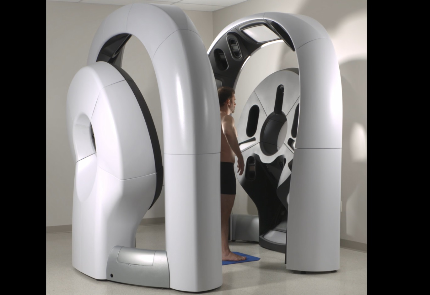
 Cutaneous neurofibromas (cNF) are the most common tumors in people with NF1, about 98% of people living with NF1 will develop these lesions. We need to learn more about how these tumors form and change over time. Knowing this information is important in order to develop better treatment options. The cutaneous neurofibroma natural history study is a research project at the Johns Hopkins Hospital aiming to learn more about how cNF form and evolve, and how they affect people who have them. In addition, this study is evaluating a new way of monitoring cNF by using a 3D camera system that can photograph the whole body in seconds. People interested in participating will have a whole-body photograph once per year for five years, complete surveys about their symptoms and receive free genetic testing of the NF1 gene.
Elana Loftspring, a young woman living with neurofibromatosis, participated in the study over the summer and shared her experience.
 In August of 2022, I had the opportunity to become a digital avatar! Earlier in the summer I had learned that Johns Hopkins University was recruiting NF patients of all ages and severities for a natural history study of cutaneous neurofibromas. I wanted to participate, but the project was in Baltimore, and I live in Houston. No problem — I discovered that my travel expenses would be reimbursed up to $700, plus I’d receive a $50 gift card! I signed up and scheduled my appointment.
I flew to Baltimore on a Thursday night. The next morning, the nurse at Johns Hopkins reviewed the consent form with me. Then she led me to a room with a very cool airport security-type of machine. I undressed and stood in the scanner for about two minutes while it took multiple pictures of me. Then I got dressed. While the nurse and I waited for the images to form, I gave a saliva sample by spitting in a tube. When that was complete, I was able to look at myself as an avatar on the computer; I could rotate it and zoom in to look closely at all my spots and bumps! I then gave a blood sample — that was optional.
The researchers on this project are using this 3D whole-body photography method to study how cutaneous neurofibromas change over a five year period. That means that I will be returning to Baltimore once a year for the next five years to do the same thing. The doctors will compare the images to see if they detect any differences in the number, color, shape or size of my neurofibromas.
Participating in this research study was quick, easy and fun. I feel good that I contributed to research that will hopefully lead to better treatments for those of us with NF1. These advances won’t happen without patients’ participation.
For information on participation, contact Mandi Johnson, Research Coordinator, at 410-502-7546 or ajohn418@jhmi.edu.
