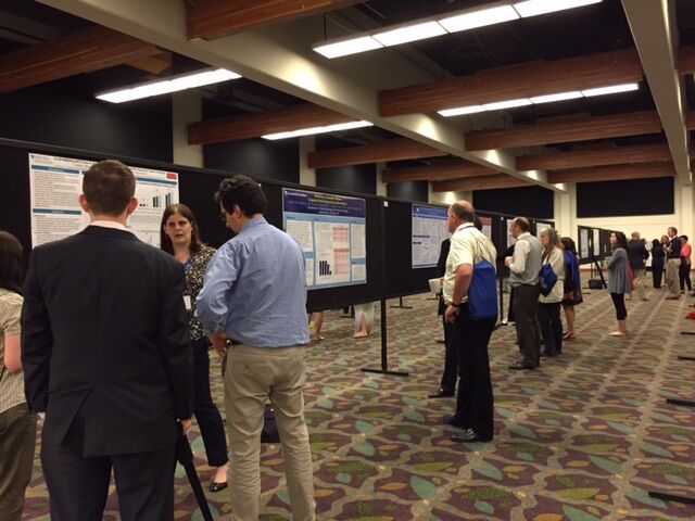
 Poster sessions are an opportunity for researchers to showcase their work in Basic and Clinical Sciences to an audience of NF researchers. A panel of judges select the top posters, and these investigators are invited to present their work in front of the full conference.
Below are the top posters at the 2015 NF Conference in Clinical and Basic Sciences.
CLINICAL SCIENCE
1) Elina Uusitalo, MSc, University of Turku, Turku, Finland
Breast cancer in NF1: incidence, survival and histopathological characteristics
2) Peter deBlank, MD, MSCE, Case Western Reserve University
The effect of carboplatin-based chemotherapy on NF1 white matter tracts associated with cognition
3) Robert A. Avery, DO, MSCE, Children’s National Health System
Automated MRI Segmention of the anterior visual pathway: establishing quantitative criteria for OPG in children with NF1
4) Kristina K. Hardy, PhD, Children’s National Medical Center/George Washington University School of Medicine
Computerized Working Memory Training for Children with Neurofibromatosis Type 1 (NF1): A Pilot Study
BASIC SCIENCE
1) Adrienne Watson, PhD, Recombinetics
Development and Characterization of a Swine Model of NF Type 1
2) Brian Kevin Stansfield, MD, Georgia Regents University
Neurofibromin Regulates Oxidative Stress and Arterial Remodeling
3) Kwangmin Choi, PhD, Cincinnati Children’s Hospital Medical Center
MicroRNA-mediated Gene Regulatory Networks in MPNST
4) Alexander Schulz MD, PhD, Leibniz Institute for Age Research, Fritz Lipmann Institute
The Importance of Nerve Microenvironment for Schwann Cell Tumors
Keep reading for the complete abstracts.
CLINICAL SCIENCE
1) Elina Uusitalo, MSc, University of Turku, Turku, Finland
Breast cancer in NF1: incidence, survival and histopathological characteristics
In order to elucidate the incidence, survival and histopathological characteristics of breast cancer in NF1, a register-based study covering whole Finland with a population of 5.4 million was carried out. The formation of the NF1 cohort has been described in Uusitalo et al. 2015 (J Invest Dermatol. 2015;135(3):904-6). The NF1 patient material was cross-referenced with Causes of Death Register, and the Finnish Cancer Registry which is population based, nationwide, and virtually comprehensive since year 1953. The results revealed a total of 44 NF1 women and one man with breast cancer. Two of the women and the male patient had two separate breast cancers. Standardized incidence ratios (SIR) and standardized mortality ratios (SMR) were calculated from cancers diagnosed between years 1987-2011 from a cohort of 1404 NF1-patients (17,947.81 person years). A total of 29 breast cancers were diagnosed during the follow-up period yielding a SIR of 3.14. SMR for breast cancer deaths was 5.26. Interestingly, in the age group of
2) Peter deBlank, MD, MSCE, Case Western Reserve University
The effect of carboplatin-based chemotherapy on NF1 white matter tracts associated with cognition
Introduction: Children with neurofibromatosis type 1 (NF1) are predisposed to low grade gliomas and are commonly treated with low-intensity carboplatin-based chemotherapy regimens thought to have no late cognitive effects. The effect of low-intensity chemotherapy on white matter integrity, separate from surgery or cranial radiation, has never been investigated. This study uses diffusion tensor imaging (DTI) to examine cerebello-thalamic tracts associated with working memory1 , and corpus callosum associated with processing speed2,3 and IQ.4 Methods: In a database of children with NF1-associated optic pathway glioma, we identified 12 subjects not exposed to chemotherapy and 12 age-matched (±1 year) controls who received chemotherapy. All tumors were restricted to the optic pathway, and no subjects received radiation or surgery. All chemotherapyexposed subjects received carboplatin/vincristine, subjects also received vinblastine (n=4), thioguanine/procarbazine/lomustine/vincristine (n=2) and bevacizumab/irinotecan (n=1). Cerebello-thalamic, thalamo-cortical and corpus callosum tracts were isolated, and a paired t-test was used to compare DTI parameters between age-matched controls. Multivariable regression was used to compare fractional anisotropy (FA) between groups, covarying for age and gradient direction set used. Results: In subjects exposed to chemotherapy, FA was decreased in cerebello-thalamic tracts (0.48±0.04 vs 0.53±0.05, p=0.03) and corpus callosum (0.65±0.04 vs 0.70±0.03, p=0.01). The effect of chemotherapy on FA remained in multivariable regression (cerebello-thalamic: -0.04±0.02, p=0.048; corpus callosum: -0.05±0.02, p=0.007). In two subjects with DTI before and after chemotherapy, FA in these tracts declined from baseline following treatment over 1 year. Discussion: Tracts associated with cognitive deficits have significantly lower FA in NF1 subjects exposed to chemotherapy. Individuals with NF1 frequently have cognitive challenges and may be more susceptible to chemotherapy-induced white matter changes. Further study is needed to examine the degree of cognitive deficit and its association with FA in patients with NF1 exposed to low-intensity chemotherapy.
3) Robert A. Avery, DO, MSCE, Children’s National Health System
Automated MRI Segmention of the anterior visual pathway: establishing quantitative criteria for OPG in children with NF1
Background: Although classification systems describe the anatomic location of optic pathway gliomas (OPGs), no quantitative criteria exist to define the presence or absence of an OPG. Since manual segmentation of MRI images is time consuming and limited to research centers, automated segmentation is necessary for accessibility. In this study, we developed and validated an automated quantitative MRI analysis algorithm of the optic nerve. Optic nerve volume and radius were compared between children with and without OPGs. Methods: Twenty-one pediatric patients (15 healthy and 6 with OPGs) with T1-weighted cube MRI sequences (~0.4 x 0.4 x 0.6mm3 ) resolution were analyzed. The optic nerve volume, average optic nerve radius and maximum optic nerve radius were calculated using: 1) manual segmentation by two operators; and 2) automated partitioned joint shape model with sparse appearance learning The mean surface distance (shortest distance between segmentation methods) and relative volume error (percent difference between volumes) were compared between segmentation methods. The volumetric values were compared between patients with and without OPGs. Results: The mean surface distance and relative volume error between manual and automated segmentation methods was 0.44±0.14 and 0.10±0.10 mm, respectively. Compared to healthy patients, OPG patients demonstrated significantly larger (P < 0.05 for all comparisons) optic nerve volume (1.002±0.522 vs. 0.342±0.098 ml), average optic nerve radius (0.800±0.293 vs. 0.401±0.050 mm) and maximum optic nerve radius (3.328±1.269 vs. 1.599±0.222 mm). Conclusions: Our automated quantitative MRI analysis algorithm of the optic nerve produced comparable results to a manual segmented method. The volumetric results comparing patients with and without OPGs were robust and well outside segmentation method variability. By developing and validating automated quantitative MRI volumetric analysis of the optic nerve in children with NF1-OPG, we will establish standardized diagnostic criteria for an OPG and provide an objective and quantitative measure of tumor growth and response to treatment.
4) Kristina K. Hardy, PhD, Children’s National Medical Center/George Washington University School of Medicine
Computerized Working Memory Training for Children with Neurofibromatosis Type 1 (NF1): A Pilot Study
Objectives: Children with NF1 have a high incidence of executive dysfunction, but few interventions have been empirically evaluated. We aimed to assess the feasibility and preliminary efficacy of a home-based, computerized cognitive training program for children with NF1 and working memory deficits.
Methods: This prospective, single-arm trial employed a pre-post design to evaluate changes in performance-based measures of attention and working memory and parent-completed ratings of executive functioning. Children meeting eligibility criteria completed training with Cogmed, a computerized working memory training program that includes phone-based coaching support over 9 weeks (25 sessions). Primary outcomes included compliance statistics (tracked automatically by the program) as well as change in attention and working memory scores. The assessment battery consisted of measures with available alternate forms and/or with few practice effects.
Results: Thirty-one (52% male; Mean age=10.9, Range=8-16) were screened for participation, with 87% showing evidence of working memory difficulties. Treatment compliance was good, with almost three-quarters of participants completing at least 80% of training sessions. There were no reported physical or psychological adverse events. Participants and their parents rated the intervention as enjoyable an appropriately challenging. Participants exhibited post-treatment improvement in attention and executive function on a number of performance-based measures, but not on the parent-rated questionnaire. Specifically, children completing training showed significantly improved sustained visual attention, verbal attention, visual working memory, and executive functioning. Preliminary resting-state MRI analyses indicated changes in functional connectivity in several regions of the brain related to attention and executive functioning.
Conclusion: Pilot data suggest that computerized training is feasible and possibly efficacious for children with NF1. Additional efficacy data will be collected through a multi-site, randomized controlled trial recently funded by the DOD. Future work should continue to evaluate functional and genetic biomarkers associated with treatment-related improvements in order to fully elucidate mechanisms of attention and executive dysfunction in this population.
BASIC SCIENCE
1) Adrienne Watson, PhD, Recombinetics
Development and Characterization of a Swine Model of NF Type 1
NF1 patients often show a variety of pathological symptoms including the development of neurofibromas, benign tumors throughout the peripheral nerves that can cause significant pain and morbidity. Secondary genetic changes, often the loss of Tumor Protein 53 (TP53), cause malignant transformation of neurofibromas in 10% of patients, which leads to the development of malignant peripheral nerve sheath tumors (MPNSTs). MPNSTs are aggressive and deadly sarcomas that are difficult to surgically resect and often are chemotherapy-resistant. There has been considerable effort in developing targeted therapies for these tumors and other NF1-associated tumors, with little success. MPNSTs remain the leading cause of death for NF1 patients. While there are several mouse models of NF1, none fully recapitulates the disease spectrum seen in the NF1 patients and preclinical work in these mice is rarely predictive of drug efficacy in humans. No drug that has emerged in NF1 mouse models has progressed past phase II clinical trials due to lack of safety and/or efficacy. Our goal is to establish a swine model of NF1 that recapitulates the phenotypic diversity of NF1 to better understand disease etiology and progression and provide a reliable preclinical model for establishing safety and efficacy studies of new therapies that will augment studies conducted in the mouse. We have used gene editing technology to create swine fibroblasts with human NF1 disease-causing alleles, and lines harboring both NF1 and TP53 mutations to model the more severe oncogenic phenotypes seen in NF1 patients. The resulting pigs are being characterized for the diagnostic criteria of NF1, peripheral nerve hyperplasia, and the development of dermal and plexiform neurofibromas, as well as paralysis, neuropathy, and mobility issues that the enlarged nerves and/or tumors may cause. We are analyzing NF1 swine for other phenotypes typically seen in NF1 patients including skeletal abnormalities, hypertension, epilepsy, optic nerve gliomas, astrocytomas, and the development of JMML. We are investigating the potential of this large animal model of NF1 for preclinical drug testing by conducting pharmacokinetic studies in healthy and NF1 swine to see if this animal model displays differential sensitivity to clinically relevant drugs. The FDA has emphasized the need for development and testing of new therapies in large animal disease models in addition to rodent models, prior to human studies. We envision this large animal model of NF1 will become a standard in evaluation of new drugs prior to Phase I clinical trials and aid in the discovery of effective treatments and cures for patients with NF1. These animals provide an ideal platform upon which to 1) study imaging technology prior to tumor development during growth and through metastasis, 2) understand tumor natural history without intervention, 3) develop minimally invasive surgical techniques and intensity modulated radiation therapy strategies, 4) perform protocol
2) Brian Kevin Stansfield, MD, Georgia Regents University
Neurofibromin Regulates Oxidative Stress and Arterial Remodeling
Persons with neurofibromatosis type 1 (NF1) are at increased risk for cardiovascular diseases including arterial stenosis. Excessive reactive oxygen species (ROS) production is linked to cardiovascular remodeling. Emerging evidence suggests that “low level” ROS induces smooth muscle cell (SMC) proliferation and may participate in the development of arterial stenosis. Utilizing primary cell culture of macrophages and SMC from Nf1 heterozygous (Nf1+/-) and WT mice, we demonstrate that neurofibromin-deficient macrophages exhibit enhanced superoxide production and Nf1+/- SMC proliferation is enhanced in the presence of low concentrations of hydrogen peroxide (H2 O2 ), while higher concentrations of H2 O2 induce Nf1+/- SMC apoptosis. Corresponding with enhanced Nf1+/- SMC proliferation, Erk is maximally activated in the presence of low concentrations of H2 O2 . To interrogate these mechanisms in vivo, we utilized a carotid artery ligation technique to induce SMC proliferation and arterial remodeling. In response to carotid artery ligation, Nf1+/- mice develop a robust neointima that is significantly reduced with daily administration of apocynin, a potent antioxidant and specific inhibitor of NADPH-oxidase 2 (NOX2) in myeloperoxidase-expressing leukocytes. Based on our previous observation that loss of Nf1 in monocytes/macrophages alone is sufficient to induce neointima formation and that NOX2 controls superoxide production via Erk and Akt activation, we intercrossed Nf1+/- mice with p47phox-/- and gp91phox-/- mice for use in our carotid artery ligation model. Both p47phox and gp91phox are necessary for NOX2 activation. In response to carotid artery ligation, genetic deletion of p47phox and gp91phox completely abrogated neointima formation in Nf1+/- mice. Quantitative analysis of the neointima from each experimental genotype shows a 50% reduction in intima area and intima/media ratio in Nf1+/- mice lacking expression of the active NOX2 complex. These data provide genetic evidence that neurofibromin regulates oxidative stress and suggests that regulation of ROS production in NF1 patients may be a viable therapeutic option for the prevention and/or treatment of cardiovascular disease in persons with NF1.
3) Kwangmin Choi, PhD, Cincinnati Children’s Hospital Medical Center
MicroRNA-mediated Gene Regulatory Networks in MPNST
Patients with Neurofibromatosis Type 1 (NF1) develop benign neurofibromas and malignant peripheral nerve sheath tumors (MPNST). One of the main hurdles in developing effective therapies for NF1-related tumors is the lack of understanding of molecular mechanisms driving tumorigenesis and malignant transformation. Whole genome mRNA expression profiling has revealed an MPNST signature of genes with differential expression relative to benign neurofibromas and normal Schwann cells. To identify the potential epigenomic control mechanisms underlying these differences in gene expression, we generated microRNA (miRNA) expression profiles from normal Schwann cells, MPNST cell lines, and MPNST primary tumors, and systematically analyzed miRNA-mediated gene regulatory networks. Both negative- and positive-correlation networks based on mRNA and miRNA expression data were generated and these correlations were further mined by integrating various genomics data, including protein-protein interaction, canonical pathways, transcription factortarget prediction, copy-number alteration, and differential DNA methylation. Based on this comprehensive data integration and mining, we identified several key miRNA-mRNA networks which may play critical roles in transformation and metastasis of MPNST, especially networks controlling the preferential activation of epithelial-mesenchymal transition (EMT) and the production of several cytokines.
4) Alexander Schulz MD, PhD, Leibniz Institute for Age Research, Fritz Lipmann Institute
The Importance of Nerve Microenvironment for Schwann Cell Tumors
Schwann cell derived tumors, referred to as schwannomas, are nerve sheath neoplasms that appear sporadically and in association with genetic syndromes such as Schwannomatosis or Neurofibromatosis type 2 (NF2). Despite their predominantly benign nature, these tumors often cause a devastating impact on patients’ life quality, not least because treatment options are often limited to surgical resection of the tumor endangering the nerve integrity and functionality. In case of NF2 disease, which is caused by the mutagenic loss of the NF2 tumor suppressor gene encoded protein merlin, the majority of patients suffer from peripheral nerve damage, clinically referred to as peripheral neuropathy. Strikingly, NF2-associated neuropathy often occurs in the absence of nerve damaging schwannomas, suggesting tumor-independent events. We have proposed a neuron-intrinsic pathogenesis of NF2-related neuropathy as merlin exerts an important role in neuronal cell types concerning axon structure maintenance. Moreover, we have identified that neuronally expressed merlin can influence Schwann cell activity in a cell-extrinsic manner via the Neuregulin-1/ErbB signaling pathway. We therefore hypothesized that the altered axon-derived cues might lead to aberrant nerve homeostasis and regeneration as well contribute to the Schwann cell tumorigenesis. We have now performed experimental nerve crush injuries on sciatic nerves of genetically modified mice in order to examine the importance of both nerve microenvironment and regeneration for schwannoma development. Strikingly, haploinsufficiency of the nf2 tumor suppressor gene when conditionally reduced gene dosage in neurons as well as their adjacent Schwann cells is sufficient to provokes schwannoma formation after nerve crush. Here, tumorlets can be found resembling affected nerves of NF2 patients. In contrast, the homozygous loss of nf2 in Schwann cells caused a macroscopic nerve thickening following nerve crush but did not meet the neuropathological criteria for schwannomas. These data address key cell intrinsic and extrinsic signaling mechanisms important for peripheral nerve repair, as well as identify a potential tumor promoting microenvironment for Schwann cells.

